
Digital radiography captures dental images using electronic sensors and computer processing rather than conventional film. These sensors register the X-ray photons and convert them into a digital file that can be viewed on a monitor in seconds. For patients, that means a faster visit and immediate visual feedback; for clinicians, it means clearer images that can be manipulated for better diagnostic insight.
Unlike older film systems that require chemical development, digital radiography transfers images directly into a secure digital record. The result is a streamlined workflow: images are available instantly, can be enhanced to reveal subtle detail, and are stored alongside other clinical data for easy retrieval. This efficiency supports more informed treatment discussions and quicker, more targeted care planning.
Digital radiography is not a single technology but a suite of tools — intraoral sensors, panoramic digital imaging, and software platforms that allow measurement, annotation, and image comparison. Together, these components form a modern imaging system that enhances communication between practitioner and patient while supporting precise diagnosis across a range of dental concerns.
One of the primary clinical advantages of digital radiography is improved image quality. Digital sensors and processing software enhance contrast and sharpness, making it easier to spot early decay, bone changes, and subtle anatomic features that might be missed on outdated film. Clinicians can zoom, adjust brightness, and compare images side-by-side to monitor progression or treatment outcomes.
Another important benefit is the ability for immediate collaboration. Digital files can be viewed on multiple screens simultaneously and shared with specialists when a referral is needed, which speeds up interdisciplinary care. This instant availability reduces wait times for second opinions and helps ensure that treatment plans are based on the most complete and current information.
Digital imaging also supports preventive dentistry. By providing clear, easy-to-interpret visuals, patients can see what the clinician sees — helping them understand why a particular treatment or follow-up is recommended. When patients can visualize cavities, bone levels, or other issues, they are better equipped to participate in decisions about their oral health.
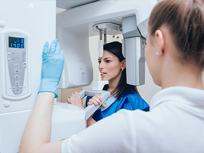
When X-rays are needed, the process in our office is designed to be comfortable, efficient, and informative. A small electronic sensor is gently positioned in the mouth to capture intraoral images. The sensor itself is thin and designed with patient comfort in mind; once the exposure is taken, the image appears on the computer monitor almost immediately for review with the clinician.
After capturing images, our clinical team uses software tools to optimize them for diagnosis — adjusting contrast, magnifying areas of interest, or measuring distances for implant planning or restorative work. Because images are stored securely in the patient's electronic chart, they become part of a comprehensive record that helps guide ongoing care and follow-up recommendations.
We place a strong emphasis on communication throughout the process. With the image displayed, your clinician will walk you through what is visible, explain clinical significance, and outline recommended next steps. Having the image on-screen during the conversation helps demystify findings and supports shared decision-making between patient and provider.
Digital radiography reduces radiation exposure compared with many traditional film-based systems because sensors are more efficient at capturing the required diagnostic information. Our practice follows professional guidelines to determine when radiographs are needed and uses modern equipment and exposure protocols to keep doses as low as reasonably achievable while maintaining diagnostic quality.
In addition to radiological safety, digital systems eliminate the need for chemical processing, making them a greener option for dental imaging. Without developer chemicals, fixer solutions, or film waste, the environmental impact of routine imaging is substantially reduced. That’s an operational improvement that aligns better with responsible practice management.
Patient privacy and secure handling of imaging data are priorities. Digital images are stored in electronic health records that employ access controls and secure backups; this helps protect sensitive information while allowing authorized members of the dental team to retrieve images when needed. When images are shared with other providers for consultation, we follow secure transfer procedures to maintain confidentiality.
Advanced imaging software also supports consistent record keeping by tagging images with date, tooth number, and relevant notes. Good documentation makes it easier to track changes over time and supports continuity of care, whether within our office or when coordinating with specialists.
Recommendations for the frequency of dental X-rays are based on individual risk factors, clinical findings, and age. New patients may receive a baseline set of images to establish a comprehensive starting point, while routine checkups might require fewer exposures. Clinicians weigh diagnostic benefit against exposure and tailor imaging schedules to each patient’s needs.
Comfort and safety measures are part of every imaging appointment. Lead aprons and thyroid collars are available when appropriate, and our staff is trained to position sensors quickly and accurately to minimize discomfort. Because images are acquired more rapidly with digital sensors, time spent with the sensor in the mouth is typically shorter than with older methods.
For pediatric patients, we take extra care to use techniques and exposure settings designed for smaller anatomies and greater sensitivity. For adults with limited mouth opening or those who experience anxiety, we adapt our approach to ensure the imaging process is as comfortable and stress-free as possible while still obtaining the information needed for excellent care.
When images indicate a need for next steps — whether watchful waiting, preventive measures, or restorative treatment — your clinician will explain the options and the rationale clearly. The goal is to use imaging as a tool that supports timely, appropriate care rather than as a routine checkbox, always prioritizing patient well-being and clinical necessity.
At Seals Family Dentistry, digital radiography is one of several technologies we use to provide clear, efficient, and patient-centered care. If you have questions about how imaging is used during your appointment or what to expect for your next visit, please contact us for more information. Our team is happy to explain our protocols and how digital X-rays contribute to precise diagnosis and thoughtful treatment planning.


Whether you're ready to schedule your first appointment, have a question about our services, or need urgent dental care, we’re just a call or click away. Our friendly team is here to make your experience simple, stress-free, and tailored to your needs.
Back to top