
An intraoral camera is a compact, pen-sized imaging device that captures high-resolution, full-color views of the inside of the mouth. Unlike traditional dental mirrors, this tool projects images in real time onto a monitor so both the clinician and the patient can see detailed views of teeth, gums, and other oral structures. The clarity and scale of these images make it easier to detect surface cracks, early decay, worn restorations, and subtle tissue changes that might be missed during a cursory visual exam.
Because it records actual photographic evidence, the intraoral camera moves routine oral exams beyond verbal descriptions and into a visual experience. This helps clinicians explain findings more precisely and patients understand their real oral condition without relying solely on dental jargon. The visual record also supports more consistent follow-up care, since clinicians can compare images from one visit to the next to track healing, progression, or the results of treatment.
In short, an intraoral camera translates what the dentist sees into clear, accurate images that improve diagnosis and communication. It’s a straightforward technology that yields outsized benefits: sharper diagnostics, better patient understanding, and more predictable treatment planning.
The detailed imagery produced by an intraoral camera enhances diagnostic accuracy by revealing fine surface details and color variations that are hard to see with the naked eye. Early-stage cavities, hairline cracks, marginal gaps in restorations, and early gum inflammation are examples of findings that become much easier to identify. When clinicians can visually confirm these conditions, they can prioritize treatments that preserve structure and prevent more extensive problems later on.
Images captured during an exam can be enlarged, paused, and reviewed in slow detail, allowing for careful assessment and collaborative decision-making among the dental team. This is especially helpful when mapping out restorative options like crowns or inlays, where the exact margins and contours matter. Having a clear photographic record also reduces uncertainty when determining whether a watchful-wait approach or a proactive intervention is the better choice.
Finally, intraoral imaging integrates well with other diagnostic tools, such as digital radiography and periodontal charting, to form a complete clinical picture. Together, these technologies let clinicians create treatment plans that are both conservative and evidence-based, minimizing surprises and focusing care on the areas that truly need attention.
One of the most practical benefits of intraoral cameras is how they transform the patient experience. Instead of relying solely on verbal explanations or abstract diagrams, patients can see the exact conditions affecting their mouths. This visual approach reduces confusion, aligns expectations, and lowers anxiety by removing much of the uncertainty that typically accompanies dental recommendations.
Seeing a clear image of a problem—such as a small area of decay or an erosion on a tooth—helps patients make informed decisions with confidence. It also supports shared decision-making: clinicians can present treatment options alongside images that illustrate what each option will address. When patients understand the “why” behind a recommendation, they are more engaged in their care and more likely to follow through with preventive steps and recommended treatments.
In addition to boosting comprehension, intraoral imaging can be a powerful educational tool for daily oral hygiene. Clinicians can show plaque accumulation, recession lines, or areas missed during brushing and then demonstrate techniques that directly address those problems. That immediate, visual feedback makes patient education both practical and memorable.
Intraoral cameras create permanent visual records that become part of the patient’s chart, improving continuity of care over time. These images are useful for monitoring progression, evaluating the outcomes of procedures, and maintaining an accurate clinical history. When a patient returns after treatment, clinicians can compare past and present images side by side to assess healing, wear, or the durability of restorations.
Those same images also make collaboration with dental specialists and dental laboratories more efficient. Clear photographs of tooth anatomy, margins, and soft tissue contours help labs fabricate restorations that fit better from the start and provide specialists with visual context before a referral appointment. This reduces back-and-forth and can shorten treatment timelines because everyone involved is working from the same, accurate visual information.
Beyond internal collaboration, recorded images can support documentation when clinical opinions or external review are necessary. The objective, dated nature of intraoral photographs helps safeguard clinical decisions and provides an objective reference point for future discussions about treatment choices or outcomes.
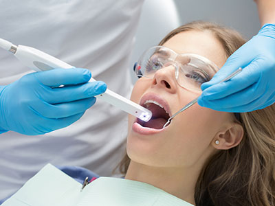
Having images taken with an intraoral camera is a quick and comfortable part of a routine dental visit. The device is small and typically used for a few minutes while the clinician captures targeted views of teeth and gums. Patients may be asked to tilt their head or bite down briefly so the clinician can photograph specific angles, but there’s no invasive procedure or recovery time associated with the imaging itself.
After the images are captured, the clinician will review them on a monitor, pointing out relevant findings and explaining possible next steps. This is an opportunity to ask questions and see exactly what the clinician is describing. The visual review typically makes treatment options and preventive strategies easier to understand and provides a clear basis for any recommended follow-up visits.
At Seals Family Dentistry, clinicians use intraoral imaging as part of a broader, patient-centered approach that emphasizes clarity and collaboration. Whether you’re in for a routine checkup or a focused follow-up, expect clear visuals to be used as a learning tool that supports precise diagnosis and a treatment plan tailored to your needs.
Summary: intraoral cameras sharpen diagnosis, improve communication, and create lasting records that support high-quality dental care. If you’d like to learn more about how this technology is used during exams or how it might benefit your next visit, please contact us for more information.

Whether you're ready to schedule your first appointment, have a question about our services, or need urgent dental care, we’re just a call or click away. Our friendly team is here to make your experience simple, stress-free, and tailored to your needs.
Back to top